What Is Patellar Instability?
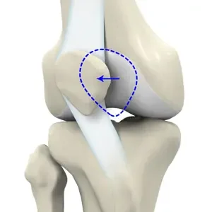
The kneecap (patella) normally glides in a V-shaped groove at the end of the thighbone (femur) as you bend and straighten your knee. Several things keep it centered in that groove: the shape of the bone itself, the muscles of the thigh, and a ligament on the inner side called the medial patellofemoral ligament, or MPFL.
Patellar instability means the kneecap slides partly or completely out of that groove. When it fully comes out it is a dislocation and when it slips out partially and returns on its own it is a subluxation. When there is a dislocation, the MPFL is usually stretched or torn in the process and the kneecap becomes more likely to slip out again.
How Patellar Instability Happens
Most dislocations happen without contact, during a pivot, twist, or awkward landing with the foot planted. A direct blow to the kneecap can also knock it out of place, but this is less common. It occurs most often in adolescents and young adults and more frequently in females.
(less common)
Underlying anatomy matters enormously. Many patients who dislocate their kneecap have one or more anatomic features that made it more likely in the first place. Identifying which of these you may have is the most important part of the evaluation because it determines both your risk of it happening again and what any surgery would need to address if needed.
Common Symptoms
- A dislocation, sometimes with a visible deformity, that may reduce on its own or needs to be put back with help
- Immediate pain and rapid swelling after the episode
- A feeling that the kneecap is slipping, shifting, or about to give way
- Apprehension or guarding with twisting, pivoting, or squatting
- Catching or grinding at the front of the knee, which may indicate cartilage damage
How It Is Diagnosed
- A detailed history of each episode, including how many and how they occurred
- Physical examination, including how the kneecap tracks, apprehension testing, and assessment of overall alignment and ligament laxity
- X-rays to assess the shape of the groove, the height of the kneecap, and any loose fragments
- MRI to evaluate the MPFL and look for cartilage damage, which is common after a dislocation
- CT in some cases to assess bony anatomy
Anatomic Risk Factors
The groove the kneecap sits in is shallow or flat rather than V-shaped.
Less bony containment to hold the kneecap in place
The kneecap sits higher than normal on the thighbone.
It enters the groove later as you bend, leaving it unprotected early in motion and more likely to dislocate
The patellar tendon attaches too far toward the outside of the shinbone.
Pulls the kneecap outward with every contraction of the thigh
A ligament on the inner side that helps keep the patella centered is stretched or torn, usually from a prior dislocation.
A main soft tissue restraint against lateral patellar movement is lost
Knock-knee alignment, or rotation of the thighbone or shinbone.
Changes the direction the kneecap is pulled during activity
Will It Happen Again?
This is the question most patients want answered, and it depends on your age, activity level, anatomy and other factors. After a first dislocation, a patient with none of the risk factors above has a lower chance of recurring. A patient with several of them has a higher chance.
This is why two people with what seems like the same injury can be given different recommendations. It is also why imaging is used to measure these features and considered as a part of a holistic evaluation. Each dislocation also carries a risk of damaging the cartilage behind the kneecap so we are mindful of preventing recurrence as much as possible.
Your Treatment Options
There is no single right answer for every patient. The decision depends on how many episodes you have had, which anatomic risk factors are present, whether cartilage was damaged, and your age and activity goals.
Nonoperative Treatment
After a first dislocation, treatment may begin with a brace and a structured rehabilitation program focused on strengthening the quadriceps and hip muscles that control how the knee tracks. The goal is a gradual return to full activity over the following weeks to months.
May be appropriate if:
- This was your first dislocation
- You have few or none of the anatomic risk factors
- There is no loose fragment of cartilage or bone in the joint
- You prefer to avoid surgery and the recovery it requires
Some first-time dislocations that are treated without surgery may not have another episode. Rehabilitation and lifestyle modifications cannot change your anatomy but can meaningfully improve the control and strength you have over the knee.
Surgical Treatment
Surgery is aimed at correcting the problems that are driving the instability, which is why the operation may not be the same for everyone.
The most common procedure is reconstruction of the MPFL, in which a new ligament is created using a tendon graft to restore the checkrein on the inner side of the knee. This is often very successful. In more serious or revision cases, when the tendon attachment sits too far to the outside or the kneecap rides too high, the attachment of the patellar tendon (the tibial tubercle) can be moved on the shinbone. In cases of severe trochlear dysplasia, the groove itself may need to be deepened. Any loose cartilage fragments are addressed at the same time. There are other options to correct anatomic risk factors as well.
Often recommended if:
- You have had recurrent dislocations or subluxations
- Your first dislocation occurred with several high-risk anatomic or demographic features present
- A fragment of cartilage or bone was knocked loose in the joint
- Instability is limiting your sport, work, or daily activities
Having anatomic risk factors does not automatically mean you need a bone procedure. Research has shown that reconstructing the MPFL alone works well for most patients, including many with mild to moderate abnormalities, and it avoids the longer recovery that comes with an osteotomy.
Recovery and Return to Sport
Recovery and Return to Activity
Without surgery:
- After a first dislocation, most people return to sport about six weeks to three months after the injury, once motion, strength and control have returned.
After surgery:
- You will use a brace and crutches early on, with weight bearing and motion advanced according to what was done.
- Recovery is longer when a bone procedure is added, because the bone must heal before full loading.
- Strengthening progresses over several months, with return to sport typically about six to seven months after MPFL reconstruction and six to eight months when a tibial tubercle osteotomy is added.
- Return to sport is guided by strength testing and confidence rather than time alone.
- Rehabilitation is the single most important factor you control!
Risks to Understand
- Instability can recur after surgery, though this is uncommon when contributing factors have been addressed.
- Knee stiffness is common early and usually improves with therapy, though occasionally it requires further treatment.
- Other risks include infection, blood clots, fracture where bone work was done, and problems related to anesthesia. This will be discussed in more detail prior to surgery.
Selected Background Sources
The guidance in this handout reflects current peer-reviewed orthopedic literature and professional society recommendations. Selected sources include:
- American Academy of Orthopaedic Surgeons. Patellar (Kneecap) Instability. OrthoInfo.
- Huntington LS, et al. Factors associated with an increased risk of recurrence after a first-time patellar dislocation: systematic review and meta-analysis. Am J Sports Med. 2020.
- Smith TO, et al. Surgical versus non-surgical interventions for treating patellar dislocation. Cochrane Database Syst Rev. 2023.
- Hurley ET, et al. Management of patellar instability: network meta-analysis of randomized controlled trials. Am J Sports Med. 2022.
- Lampros RE, Tanaka MJ. Return to play considerations after patellar instability. Curr Rev Musculoskelet Med. 2022.
- Platt BN, et al. Return to sport after MPFL reconstruction: systematic review and meta-analysis. Am J Sports Med. 2022.
- Schneider DK, et al. Outcomes after isolated MPFL reconstruction: systematic review and meta-analysis. Am J Sports Med. 2016.
- Erickson BJ, et al. Isolated MPFL reconstruction regardless of tibial tubercle-trochlear groove distance and patellar height. Am J Sports Med. 2019.
- Dennis ER, et al. Isolated MPFL reconstruction regardless of TT-TG distance and patellar height: minimum 5-year outcomes. Am J Sports Med. 2024.
- Dandu N, et al. Trochlear bump and patella alta predict recurrence after MPFL reconstruction. Am J Sports Med. 2025.
- Ryan PC, et al. Fear of reinjury and psychological factors in return to sport after MPFL reconstruction. Arthroscopy. 2025.
- Parmar R, Keane A, Tummala SV, et al. Diagnostic arthroscopy for chondral lesions missed by MRI in pediatric MPFL reconstruction. Orthop J Sports Med. 2025.
This handout is for education and does not replace the discussion with your surgeon. Ask Dr. Tummala about anything that applies to your specific knee.
Handouts for this condition
- Condition guideThe printable version of this pageOpen PDF
- Physical therapy protocolNonoperative plan to bring to your therapistOpen PDF
If you are having surgery
You will receive a guide to your specific operation at your visit. Read what to expect from MPFL Reconstruction, including the instructions and therapy protocols for after surgery. The Preparing for Surgery Booklet is on the Having surgery page.