What Is Knee Arthritis?
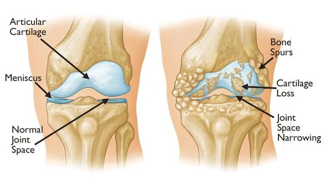
The ends of the bones in your knee are covered by a smooth layer of cartilage that lets the joint glide with almost no friction. In arthritis, a combination of inflammation and degeneration causes that cartilage to wear over time. As it thins, the joint becomes stiffer and more painful, bone spurs form, and the joint may become swollen and inflamed.
The knee has three separate compartments: the inner side, the outer side, and the area behind the kneecap. Arthritis can affect one, two, or all three. Which compartments are involved matters because it changes what treatments can be used. There is no way to regrow cartilage currently, so the goal of treatment is to control symptoms and keep you as active as possible.
How Knee Arthritis Develops
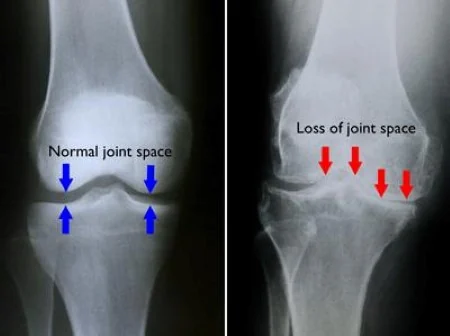
Most knee arthritis develops gradually over years as cartilage wears down. It can also follow an injury, particularly a previous fracture, ligament tear, or removal of meniscus tissue, in which case it may appear at a considerably younger age. Less commonly it results from an inflammatory condition such as rheumatoid arthritis.
Several factors increase risk including age, body weight, previous knee injury or surgery, family history, and the alignment of your leg. Alignment matters because a bow-legged or knock-kneed limb concentrates load on one side of the joint.
Common Symptoms
- Aching pain that worsens with activity and eases with rest
- Stiffness after sitting or first thing in the morning that loosens as you move throughout the day
- Swelling that comes and goes, often after more activity than usual
- Grinding, clicking, or a sense of the knee catching
- Difficulty with stairs, squatting, or getting up from a low chair
- Pain that may worsen with weather changes or at night as arthritis progresses
How It Is Diagnosed
- A detailed history of your symptoms, prior injuries, and what limits you day to day.
- Physical examination, including motion, alignment, swelling and where the tenderness is.
- Standing X-rays, which show joint space narrowing, bone spurs, and which compartments are involved.
- Alignment views in some cases, particularly when an osteotomy is being considered.
- MRI is often not needed but can be used in select cases. When arthritis is clear on X-ray, it rarely changes the plan.
Where the Arthritis Can Be
Wear on the inner side of the knee. This is the most common pattern.
Often associated with bow-legged alignment. May be treatable with an osteotomy, partial replacement or total knee replacement depending on the extent of arthritis.
Wear on the outer side of the knee.
Often associated with knock-kneed alignment. Also potentially treatable with an osteotomy, partial knee replacement or total knee replacement.
Wear behind the kneecap.
Causes pain with stairs, squatting, and prolonged sitting more than with walking.
Wear affecting more than one area of the knee.
Joint-preserving options are more limited, and replacement becomes more likely if symptoms are severe.
What Helps The Most
Two things consistently do more for arthritis symptoms than anything else and both are within your control. The first is regular exercise, particularly strengthening the muscles around the knee. Strong muscles absorb load that would otherwise go through the joint. The second is weight management, because the force through the knee with every step is a multiple of body weight, so even a small reduction meaningfully unloads the joint.
Diet plays a role as well. An anti-inflammatory eating pattern with less processed food and sugar supports both weight management and joint health. Glucosamine and chondroitin are widely used but have not been shown to slow or reverse arthritis. Biologic injections such as platelet-rich plasma and stem cells are reasonable options to discuss, though the evidence is still developing. Opioids are not effective for arthritis pain and are best avoided.
Your Treatment Options
Treatment is a stepwise process. Almost everyone starts with nonoperative care, and many patients stay there successfully for years. Surgery is considered when symptoms are no longer controlled and quality of life is meaningfully affected.
Nonoperative Treatment
Exercise and physical therapy
This is the foundation of treatment. Strengthening the muscles around the knee reduces the load the joint has to absorb.
Pain management and inflammation control
Acetaminophen and anti-inflammatory medication by mouth or as a topical gel can be used. When medication is not enough, injections into the joint are the next step. Corticosteroid injections are the most established. Hyaluronic acid, platelet-rich plasma, and stem cell injections are also used though the evidence supporting them is less consistent.
Lifestyle modification
Weight management when relevant and shifting toward lower-impact activity such as cycling, swimming, and walking are helpful approaches.
Support
An unloader brace may help when arthritis is confined to one side.
If a knee replacement is being planned, it is generally recommended to wait at least three months after any injection into the joint before surgery. Injecting closer to the operation is associated with a higher risk of infection.
Surgical Treatment
Surgical options depend on your age, which compartments are involved, and your alignment. Arthroscopy, or cleaning out the joint, is generally not recommended for arthritis alone. It has not been shown to provide lasting benefit over exercise therapy even when a degenerative meniscus tear is seen on MRI without mechanical symptoms.
For younger, active patients with arthritis limited to one compartment and malalignment, an osteotomy can be considered to shift weight away from the worn side of the knee onto the healthier side. This preserves your own joint and does not carry the activity restrictions of a replacement, at the cost of a longer recovery while the bone heals.
When arthritis is advanced and affecting quality of life, joint replacement is highly effective at relieving pain, either partial or total replacement depending on the pattern. Dr. Tummala will discuss this when that is the right step.
Often recommended if:
- Pain persists despite exercise, weight management, medication, and injections
- Pain is waking you at night or severely limiting your daily activities
- You are younger with one-sided arthritis and malalignment, where an osteotomy may preserve your own knee
- X-rays show advanced arthritis that matches your symptoms
Timing matters. An osteotomy is generally best suited to younger patients before arthritis becomes widespread, while a replacement is usually delayed as long as symptoms allow.
What to Expect
Living With Knee Arthritis
- Arthritis symptoms typically fluctuate. Good stretches and flares are both normal, and a flare does not mean the arthritis has suddenly worsened.
- Low-impact activity such as using a stationary bike is encouraged. Motion helps a joint with arthritis.
- After an osteotomy, weight bearing is restricted while the bone heals, with return to full activity over several months.
- After a replacement, most patients return to everyday activities and low-impact recreation, though high intensity running and impact sports are usually discouraged.
- Staying active and maintaining strength is the single most important factor you control!
Risks to Understand
- Repeated corticosteroid injections into the same joint are generally limited in number. They lose efficacy over time and may increase the risk of cartilage injury.
- Other risks of surgery include infection, blood clots, stiffness, and problems related to anesthesia. This will be discussed in more detail prior to surgery.
Selected Background Sources
Reflects current peer-reviewed literature and professional society recommendations:
- American Academy of Orthopaedic Surgeons. Clinical Practice Guideline: Management of Osteoarthritis of the Knee (Non-Arthroplasty).
- Kise NJ, et al. Exercise therapy versus arthroscopic partial meniscectomy for degenerative meniscal tear: randomized controlled trial with two-year follow-up. BMJ. 2016.
- Berg B, et al. Arthroscopic partial meniscectomy versus exercise therapy for degenerative meniscal tears: 10-year follow-up of the OMEX randomised controlled trial. Br J Sports Med. 2025.
- Thorlund JB, et al. Arthroscopic surgery for degenerative knee: systematic review and meta-analysis of benefits and harms. BMJ. 2015.
- Brouwer RW, et al. Osteotomy for treating knee osteoarthritis. Cochrane Database Syst Rev. 2014.
This handout is for education and does not replace the discussion with your surgeon.
Handouts for this condition
- Condition guideThe printable version of this pageOpen PDF
If you are having surgery
You will receive a guide to your specific operation at your visit. Read what to expect from High Tibial Osteotomy, including the instructions and therapy protocols for after surgery. The Preparing for Surgery Booklet is on the Having surgery page.