What Is Articular Cartilage?
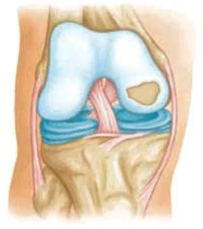
Articular cartilage is the smooth, white surface covering the ends of the bones in your joints. It allows joints to glide with almost no friction and spreads load across the surface. Unlike most tissues in the body, it has a poor blood supply limiting its ability to heal after serious injury.
A cartilage injury means a part of that surface has been damaged, much like a pothole in an otherwise smooth road. When the bone underneath is damaged as well, it is called an osteochondral injury. This is different from arthritis, where the surface wears across a broad area though it is a spectrum of disease. A focal defect in an otherwise healthy knee is potentially repairable, which is why distinguishing the two matters.
How Cartilage Injuries Happen
Some cartilage injuries are traumatic from a direct impact, a twisting injury, or a kneecap dislocation. They frequently occur alongside other injuries such as ligament tears, meniscus injuries and even fractures. Others develop slowly where the joint has been overloaded for a long time often because of malalignment or loss of meniscus tissue.
Cartilage itself has no nerve endings, so the damaged surface is not what hurts. Pain comes from irritation of the bone underneath and the lining of the joint. This is part of why some defects cause considerable symptoms while others, sometimes found incidentally during surgery for another problem, cause none at all and may not need treatment.
Common Symptoms
- Pain localized to one area of the knee, often worse with loading that part of the joint
- Swelling that recurs after activity
- Catching, clicking, or a sense of something moving inside the knee
- Locking or giving way, which can occur if a fragment has become loose
- Symptoms that are often intermittent rather than constant
How It Is Diagnosed
- A detailed history of your symptoms and any prior injury or surgery.
- Physical examination to localize the pain and assess alignment, ligament, and meniscus status.
- X-rays including alignment views, which show the bone and overall alignment of the limb.
- MRI to help identify the defect and assess its size, depth, and whether the bone beneath is involved.
- MRI can underestimate the true size of a defect or miss defects, so the final plan may need to be determined by arthroscopy.
Cartilage Restoration Options
Loose or unstable cartilage edges are smoothed to reduce mechanical irritation.
Often the first surgical step for smaller unstable flaps, or as an adjunct to other procedures
Small cylinders of cartilage and bone are taken from a low-load area of your own knee and transferred into the defect.
Smaller defects, using your own tissue in a single operation
A matched piece of donor cartilage with its underlying bone is shaped and placed into the defect.
Larger defects, and defects where the bone beneath is also damaged
Your own cartilage cells are harvested, grown in a laboratory, and implanted on a membrane in a second operation.
Medium to large defects where the bone underneath is intact
The Knee Has to Be Set Up to Succeed
Cartilage repair does not happen in isolation and there are multiple factors that determine its success. Notably, the alignment of your leg, an intact meniscus to spread load, and stable ligaments are all critical. If there are major deficiencies in these categories, the repaired area may be more likely to fail.
This is why an evaluation for cartilage surgery looks at the whole knee rather than just the defect, and why a realignment osteotomy, meniscus transplant, or ligament reconstruction is sometimes recommended at the same time or beforehand. Addressing these issues gives cartilage restoration a chance to work.
Your Treatment Options
Not every cartilage defect needs surgery. The decision depends on your symptoms, the size and location of the defect, your age and activity level, and the overall condition of the knee.
Nonoperative Treatment
Physical therapy to strengthen the muscles around the knee, activity modification to reduce impact loading, and anti-inflammatory medication can be considered. An injection may help with symptoms. An unloader brace can reduce load on the affected side if indicated.
May be appropriate if:
- The defect was found incidentally and is not causing symptoms
- Your symptoms are mild or intermittent
- The defect is small and stable
- You prefer to avoid surgery and the recovery it requires
Nonoperative treatment works for many patients with a cartilage lesion who have manageable symptoms and they may never need surgery for it.
Surgical Treatment
The goal of cartilage restoration is to fill the defect with durable tissue that can bear load. Which procedure is chosen depends on the size and depth of the defect, whether the bone underneath is involved, its location, and your age and activity level.
Some procedures are done in a single operation others require multiple stages.
A diagnostic staging arthroscopy may be performed first to confirm the size and character of the defect directly under camera visualization to ensure the correct treatment plan for you.
Often recommended if:
- You have a symptomatic defect that has not responded to nonoperative care
- A fragment has become loose in the joint or the knee is locking
- You are young and active with an isolated defect in an otherwise healthy knee
- The defect is large enough that it is unlikely to become asymptomatic on its own
Cartilage restoration is generally not appropriate once arthritis has become widespread. These procedures work best for isolated defects in an otherwise healthy knee.
What to Expect After Surgery
Recovery and Return to Activity
- Recovery after cartilage restoration is long. Weight bearing is usually restricted for several weeks to protect the repair while it integrates.
- Motion often begins early, sometimes with a machine that moves the knee passively, because motion helps cartilage heal.
- Return to running and impact activity is typically many months out, and return to sport is often around nine months or more.
- The repaired tissue continues to mature well beyond the point where the knee feels good, which is why restrictions are followed even when you feel ready.
- Rehabilitation is the single most important factor you control!
Risks to Understand
- The repair can fail, and a second procedure is sometimes needed.
- Knee stiffness is common early and usually improves with therapy, though occasionally it requires further treatment.
- Procedures using donor tissue depend on graft availability which can affect the timing of surgery.
- Cartilage restoration does not guarantee you will avoid arthritis later and the goal is to delay progression and improve symptoms.
- Other risks include infection, blood clots, and problems related to anesthesia. This will be discussed in more detail prior to surgery.
Selected Background Sources
Reflects current peer-reviewed literature and professional society recommendations:
- Wang AS, et al. Minimum 10-year outcomes of matrix-induced autologous chondrocyte implantation in the knee: systematic review. Am J Sports Med. 2024.
- Brittberg M, et al. Five-year follow-up of the SUMMIT randomized trial of matrix-applied chondrocyte implantation. Am J Sports Med. 2018.
- Familiari F, et al. Clinical outcomes and failure rates of osteochondral allograft transplantation in the knee: systematic review. Am J Sports Med. 2018.
- Pareek A, et al. Long-term outcomes after osteochondral autograft transfer: systematic review. Arthroscopy. 2016.
- Salem HS, et al. Staging arthroscopy before cartilage restoration surgery. J Knee Surg.
- Gomoll AH, et al. MRI underestimates articular cartilage defect size. Cartilage. 2011.
- Flanigan DC, et al. Prevalence of chondral defects in athletes’ knees: systematic review. Med Sci Sports Exerc. 2010.
This handout is for education and does not replace the discussion with your surgeon.
Handouts for this condition
- Condition guideThe printable version of this pageOpen PDF
If you are having surgery
You will receive a guide to your specific operation at your visit. Read what to expect from Cartilage Restoration and High Tibial Osteotomy, including the instructions and therapy protocols for after surgery. The Preparing for Surgery Booklet is on the Having surgery page.